
Address
Königinstrasse 10
80539 München, Germany
Google-Maps
5H2P+M7 München
Application Spotlight
Cells & Tissue
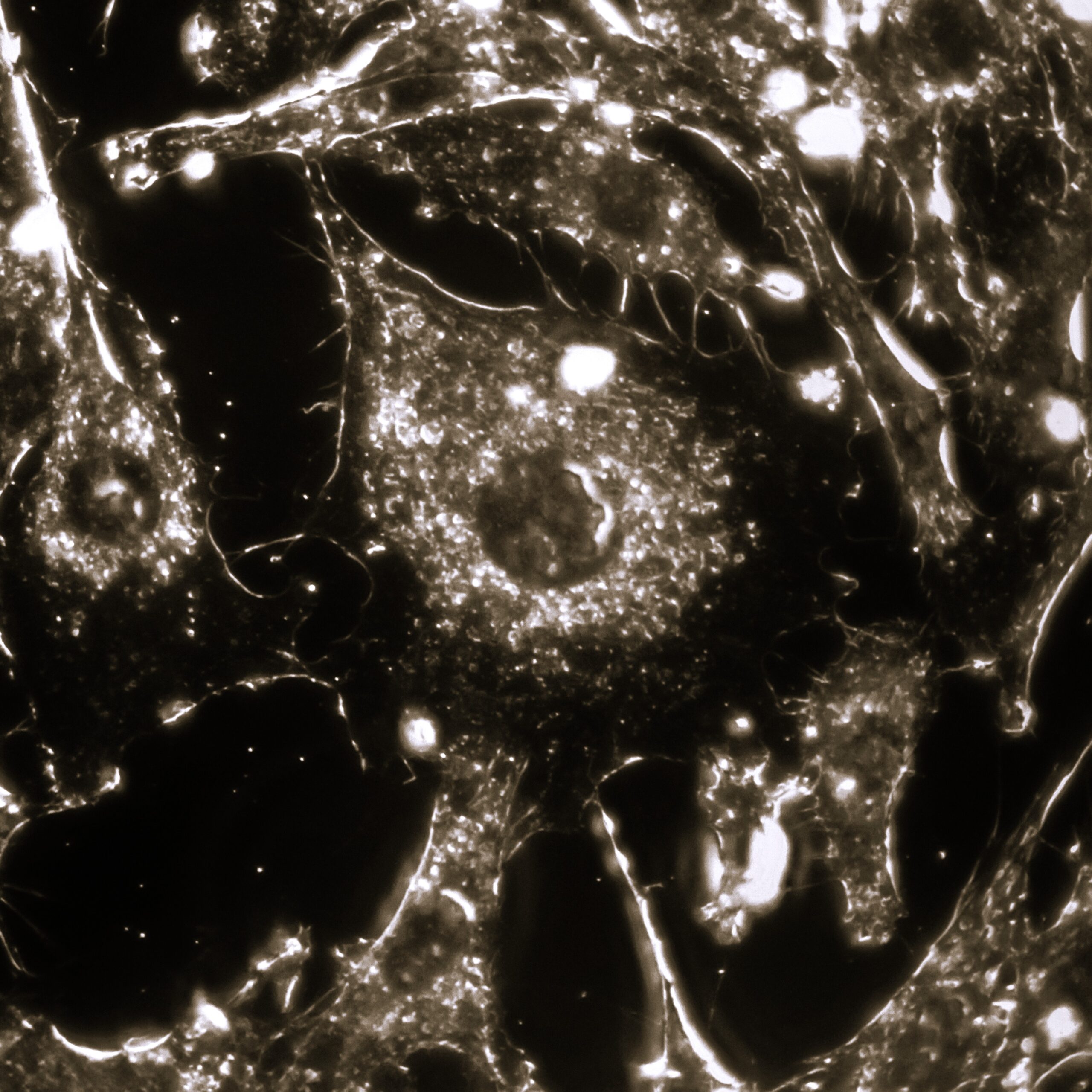
Our microscope lets you see unlabeled biomolecules and vesicles in living systems with nanometer precision and micro‑ to millisecond dynamics—without photobleaching, phototoxicity, or labeling artifacts. That means you can interrogate real biological behavior, at the native mass and in the native environment.
Frequently Asked Questions (FAQs)
In Lifescience when working with cells and tissue, your microscope is required to operate
- Label‑free: No dyes, no tags, no perturbation.
- Ultra‑fast: Tracks rapid transport and binding events in real time.
- Ultra‑precise: Nanometer localization and sub‑nanometer axial sensitivity.
- Gentle on samples: Minimal light dose; ideal for long time‑lapse studies.
- Versatile: From single proteins at membranes to cargo flow deep in tissue slices.
We detect tiny intensity changes of photons emerging from a nanoscale object (protein, virus, vesicle) and its interaction with a background from the coverslip or tissue interface. The contrast scales with particle effective mass, enabling direct detection and quantitative tracking of unlabeled species against complex cellular or extracellular backgrounds..
Worth noting: The system also avoids the fading (photobleaching) and other limitations of conventional fluorescence microscopy.
- Membrane processes: Single‑protein diffusion, receptor nano‑clustering, endo/exocytosis, virus docking & entry.
- Intracellular transport: Vesicles and organelle precursors along cytoskeleton with sub‑ms temporal resolution.
- Cell–matrix & cell–cell interactions: Adhesion dynamics, immune synapse formation, ECM remodeling.
- Extracellular vesicles (EVs): Size‑dependent scattering contrast for label‑free EV counting and tracking.
- Tissue slices: Perfused brain, cardiac, or tumor slices—follow particle transport and drug–target engagement in native microenvironments.
- Cell therapy & gene delivery: Vector binding, entry, and trafficking without fluorescent modification.
- Immuno‑oncology: Immune synapse dynamics, checkpoint clustering (native receptors).
- Neuroscience: Axonal transport and synaptic vesicle dynamics in slices; activity‑dependent trafficking.
- Biomaterials: Protein corona formation and matrix remodeling on implantable surfaces—in situ.
- Diagnostics: Label‑free particle characterization (EVs, pathogens) as front‑end to molecular ID.
- No labels = true biophysics: Avoids steric effects, altered kinetics, and photobleaching.
- Speed + SNR: The system delivers high SNR at high frame rates (fast trafficking, short‑lived binding).
- Mass contrast: Signal scales with particle polarizability → relative mass readout and heterogeneity mapping.
- Depth & complexity: Works on live cells, organoids, and tissue slices where labeling is challenging or disruptive.
- Easy correlation: Combine with AFM, EM, patch‑clamp, or microfluidics to link structure, mechanics, and function.